Dosimetry for Patients
Dose to patients can be estimated in a number of ways depending on the intended purpose and the accuracy required. The estimated dose is given accordingly as dose indices, absorbed dose, or effective dose. These indicators of dose can be measured using appropriate equipment and phantoms, or calculated, similarly with appropriate tools.
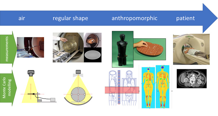
Air kerma is used as a measure of the radiation output of a diagnostic imaging system, and thus can give some indication of the effect of changing protocol settings. Absorbed dose can be estimated using regular shaped and homogenous phantoms, or detailed anthropomorphic phantoms containing simulated organs enabling organ dose to be estimated.
By taking the organ doses from an anthropomorphic phantom, multiplying each organ dose by the appropriate ICRP tissue weighting factor, and then summing over all organs, an effective dose is calculated. Thus, the estimation of dose is taken on a path towards patient specific dosimetry.
The Medical Dosimetry Group at UKHSA performs numerical dose calculation by using Monte Carlo dose calculations. Monte Carlo calculations are calculations using statistical interactions of individual photons (X-rays in diagnostic radiology), to estimate organ doses from an X-ray source. The primary work of the group in this area is currently focussed on computed tomography.
To undertake these calculations the scanner, the patient and the X-ray interactions must all be modelled. Specifically, the numerical MIRD-like phantoms and ICRP voxel phantoms are used, together with the modelling of the physical characteristics of a range of computed tomography scanner models, based on information supplied by the manufacturers of these systems. Different protocol, tube voltage and filtration options, are also modelled.
Radiation Output to Patient Specific Dosimetry
|
Details of previous patient dosimetry work carried out by MDG
|
Read more |
|
Details of the latest patient dosimetry work being carried out by MDG
|
Read more |
|
A free calculator to estimate patient dose
|
Read more |